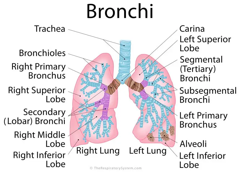

The inner mucosal layer has a lining of ciliated pseudostratified columnar epithelium with goblet cells. The posterior aspect of the trachea is adjacent to the anterior esophagus. A narrow thin membrane connects each of the tracheal rings. The trachea is 16 to 20 C-shaped cartilage rings stacked one on top of another. It is much broader in the proximal area than the distal segment. The tracheal diameter is about 24 to 26 mm in adult males and 22 to 24 mm in females. This junction point is called the carina. The trachea is a midline structure and lies just anterior the esophagus.Īfter it originates from the larynx, the trachea divides into the left and right mainstem bronchi. The primary function of the trachea is to allow passage of inspired and expired air into and out of the lung. Common asthma triggers.The trachea originates at the inferior edge of the larynx and connects to the left and main stem bronchus. Lung procedures, tests & treatments.Ĭenters for Disease Control and Prevention. Morphometric analysis of explant lungs in cystic fibrosis. Bronchiolitis obliterans.īoon M, Verleden SE, Bosch B, et al. National Center for Advancing Translational Sciences/National Institutes of Health. Respiratory syncytial virus infection and bronchiolitis.

Overview and challenges of bronchiolar disorders. Swaminathan AC, Carney JM, Tailor TD, Palmer SM. The impact of cold on the respiratory tract and its consequences to respiratory health. The physiological basis of pulmonary gas exchange: implications for clinical interpretation of arterial blood gases. The role and importance of club cells (Clara cells) in the pathogenesis of some respiratory diseases. Rokicki W, Rokicki M, Wojtacha J, Dżeljijli A. Elastin in lung development and disease pathogenesis. A unique cellular organization of human distal airways and its disarray in chronic obstructive pulmonary disease. The comparison of the lengths and diameters of main bronchi measured from two-dimensional and three-dimensional images in the same patients.


 0 kommentar(er)
